Navigating Breast Health: Understanding Breast Calcifications
Every woman is aware of the significance of maintaining breast health, and routine screenings are a cornerstone of identifying any issues at an early stage. Among the findings that can appear on mammograms, breast calcifications are common yet can evoke concern and raise questions for many women. Understanding the nature of breast calcifications, including their potential causes, prevalence, and the importance of follow-up examinations, is essential for a comprehensive understanding of breast health and enables women to make well-informed decisions.
What Are Breast Calcifications?
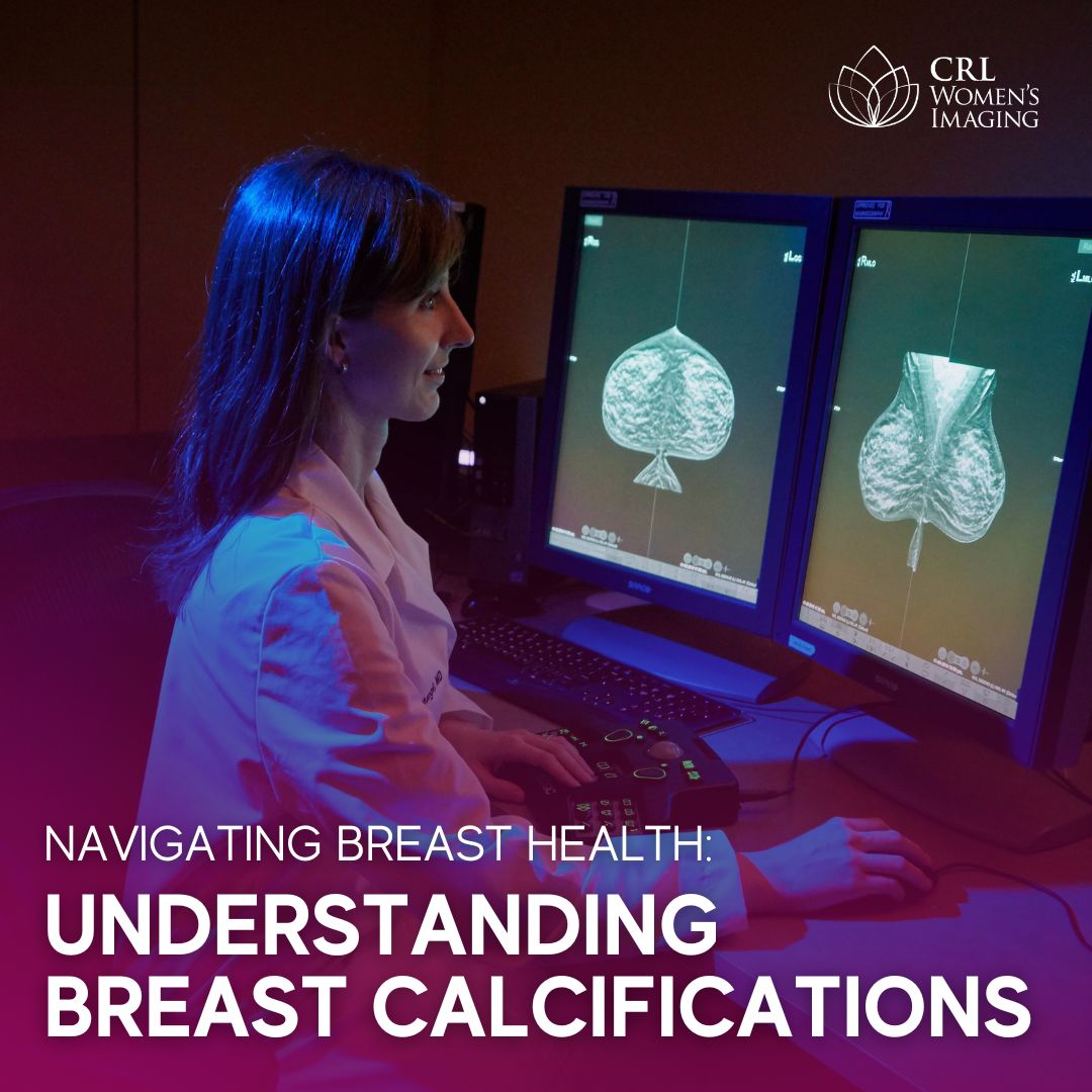
Breast calcifications are tiny deposits of calcium that can develop within breast tissue. These small specks are painless and imperceptible to touch. One might wonder, are these related to the calcium we consume in our diets? The answer is no; breast calcifications are not associated with dietary intake of calcium. Rather, breast calcifications act as indicators, pointing to a process going on inside the breast tissue. They are frequently detected during routine mammograms, showing up as bright white dots on the imaging.
How Common Are Breast Calcifications?
Approximately half of all women may experience breast calcifications at some point in their lives. They become more prevalent with age, especially among women over 50. The good news is that in the majority of cases, breast calcifications are benign and non-cancerous. However, due to a small chance of malignant calcifications, additional testing might be recommended to rule out any potential concerns.
Possible Causes of Breast Calcifications
While the exact cause of breast calcifications remains undetermined, a number of possible explanations are commonly acknowledged by medical experts. These include normal aging, fibrocystic changes in the breast, aging arteries, breast cysts, fibroadenomas (benign growths or tumors), previous breast injuries or infections, prior breast surgeries or cancer treatments, as well as cancer and atypical cells within the breast.
Types of Breast Calcifications
Breast calcifications are categorized into two types based on their morphology and distribution: benign and suspicious.
- Benign calcifications are larger, round, punctate, or rod-like and are often distributed throughout the breast.
- Suspicious calcifications start to form in a group, line, or segment, indicating they may be forming within a milk duct. The morphology is often irregular or branching.
Additional magnified views of the breast help determine the morphology and distribution.
The Importance of Follow-up Exams
When breast calcifications are detected, follow-up exams, including diagnostic mammograms, may be recommended. Diagnostic mammograms provide more detailed images of specific areas of concern, allowing radiologists to thoroughly assess the size, shape, and distribution of the calcifications.
If the calcifications appear benign, no further action may be needed. However, if they appear indeterminate or suspicious, additional tests, such as a stereotactic-guided breast biopsy, may be recommended. This minimally invasive procedure uses mammography for guidance to extract a small tissue sample for analysis. This step ensures a more accurate diagnosis and guides any necessary next steps.
Empowering Women with Knowledge
Understanding breast calcifications and their implications empowers women to take charge of their breast health. Regular mammograms and prompt follow-up examinations, if necessary, play a crucial role in early detection and effective management. Knowledge is a powerful tool, and being informed allows women to make confident and informed choices about their health.
Navigating breast health involves not only routine screenings but also awareness and understanding of findings like breast calcifications. By fostering a proactive approach and staying informed, women can confidently prioritize their well-being and continue on the path to lifelong breast health.
About CRL Women’s Imaging
CRL Women’s Imaging is a leader in outpatient imaging and designated as Breast Center of Excellence by the American College of Radiology (ACR). Our team of dedicated, board-certified breast imagers with broad expertise and a genuine interest in breast imaging and our knowledgeable technologists certified in mammography and ultrasound are committed to provide our patients with the high-quality compassionate care they can trust.
“Early detection of breast cancer saves lives. And with the tools of 3D mammography/tomosynthesis and supplemental screening breast ultrasound we are better equipped than ever to positively impact women’s health.” – Jillian Karow, MD, Medical Director, CRL Women’s Imaging