Breast Cancer Staging: What the Stages Mean and Why Early Detection Matters
Breast cancer staging helps describe how large a breast cancer is and whether it has spread—information your care team uses to plan treatment. In this guide, we explain breast cancer stages 0 through IV, the role imaging plays in staging, and why regular screening mammograms are a key part of early detection.
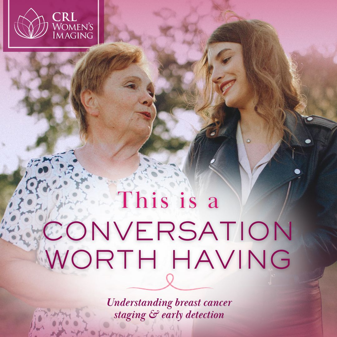
Some of the most important conversations we never have are about health. Not because we don’t care, but because we don’t know where to start — or we keep waiting for the right moment. The truth is, the right moment is usually right now, and the conversation worth having is about breast health — yours, and the people you love.
Breast cancer doesn’t follow a single script. According to the National Breast Cancer Foundation, only 5–10% of individuals diagnosed with breast cancer have a family history of the disease — meaning the vast majority of diagnoses occur in people with no known family history at all. It’s one of the most important reasons annual screening matters for everyone, regardless of background or genetics. And if the conversation ever shifts from prevention to diagnosis, for yourself or someone you love, understanding what comes next can make the difference between feeling lost and feeling prepared.
Breast cancer staging is one of those topics that sounds intimidating but doesn’t have to be. Once you understand the basics, it becomes a framework for making sense of a diagnosis — not a source of fear. Here’s what it means, why it matters, and how this knowledge can help you support the people you love, or yourself.
Breast Cancer Staging Explained: What the Stages Mean
When breast cancer is diagnosed, the next question is always: how much is there, and where is it? Staging answers that question. It gives doctors and patients a common language for describing what’s happening in the body so that treatment decisions can be made with clarity and confidence.
According to the National Cancer Institute, breast cancer stage is based on several factors: the size and location of the primary tumor, whether the cancer has spread to nearby lymph nodes, whether it has spread to other parts of the body, the grade of the tumor (how the cancer cells look under a microscope), and the status of certain biological markers — including estrogen receptor (ER), progesterone receptor (PR), and HER2. These factors are combined to assign a stage from 0 through IV.
Stage 0 means the cancer cells are present but have not spread beyond their original location — a noninvasive cancer also called carcinoma in situ. Stage I describes a small invasive cancer with limited or no lymph node involvement. Stage II means the tumor is larger and may have spread to a small number of nearby lymph nodes. Stage III indicates the cancer has spread more extensively to lymph nodes or to tissue near the breast. Stage IV means the cancer has spread to other parts of the body, most often the bones, lungs, liver, or brain.
It’s worth saying plainly: stage is a description, not a verdict. It guides treatment decisions and helps the care team understand the full picture, but it doesn’t determine any individual person’s outcome. Medicine has advanced significantly, and women are living longer and doing better across all stages than statistics from even a decade ago reflect.
“The imaging we provide is often the first step in understanding what’s happening — it’s what gives the entire care team the information they need to move forward. When patients understand what we’re looking for and why, it makes every part of that process less frightening and more actionable.”
— Dr. Jillian Karow, Medical Director, CRL Women’s Imaging
How Mammogram, Ultrasound, and MRI Help with Breast Cancer Staging
Radiology is central to staging — and most people don’t realize how much of this work happens before a treatment plan is ever formed. Imaging doesn’t just find cancer; it helps map it.
Mammography is typically where the initial finding is made. It can identify the primary tumor and show whether there appear to be multiple areas of concern within the same breast. Ultrasound provides additional detail: it’s particularly useful for evaluating the lymph nodes under the arm, assessing the characteristics of a mass, and guiding biopsies. MRI is often brought in for surgical planning — it gives a detailed view of the extent of disease, helps evaluate the opposite breast, and is especially valuable when surgeons need to understand the full picture before deciding on next steps.
According to the American College of Radiology’s Appropriateness Criteria for imaging of invasive breast cancer, selecting the right imaging at each point in care is essential — not only to identify what’s there, but to ensure the process is efficient and focused. Each imaging tool tells a different part of the story. Together, they give the care team the clearest possible picture of what they’re working with, which directly shapes treatment options.
If You’ve Been Diagnosed (or Someone You Love Has): What to Expect Next
If a family member or close friend receives a breast cancer diagnosis, your first instinct might be to research everything immediately — or to feel so overwhelmed that you don’t know where to start. Understanding staging can help you find a middle ground: enough knowledge to be present and helpful, without carrying the weight of trying to understand everything at once.
When a concerning finding is identified through imaging, it helps to know that what comes next takes time. Additional imaging is often ordered to get a clearer picture. A biopsy may be needed to confirm what the images show. Those results go to the referring physician, who works with the broader care team to determine next steps. This isn’t delay — it’s thoroughness, and it’s what good, careful medicine looks like.
If someone close to you is navigating this, the most helpful thing you can do is encourage them to write down questions before each appointment, ask for clarification when something isn’t clear, and bring a second set of ears. Information absorbs differently when you’re anxious, and having someone alongside can make a real difference. An early stage doesn’t minimize the experience — it is still cancer, and it still deserves full attention. But it does often mean a broader range of treatment options. An advanced stage is not a closed door. It’s a different set of medical decisions, and people navigate treatment and live meaningful lives at every stage.
The Mother-Daughter Conversation Worth Having
The conversations we have — or haven’t had — with the women in our families matter more than most of us realize. Your mother’s breast health history is genuinely relevant to your own. Whether she had a biopsy, when she started screening, whether any findings were ever flagged — these details matter to your risk assessment and, potentially, to when your own screening should begin.
If you have daughters, consider what you’d want them to know. While most breast cancers occur in people with no family history, a first-degree relative with breast cancer — a mother, sister, or daughter — can influence when screening is recommended to start. Sharing what you know about your own health history is useful information, even if that history is unremarkable. The conversation itself models the habit of paying attention.
If the topic feels awkward, it doesn’t have to be a formal sit-down. It can start with something simple: “I just scheduled my mammogram — have you been keeping up with yours?” Or: “I’ve been thinking about our family health history and wanted to make sure you know what I know.” It’s not a heavy conversation. It’s a caring one.
What to Do with This Knowledge
If you’re currently in good health and up to date on screening, the takeaway is straightforward: keep going. CRL Women’s Imaging follows the guidelines of the American College of Radiology (ACR) and the Society of Breast Imaging (SBI), which recommend annual mammography screening beginning at age 40 for women at average risk. Research shows this approach provides the greatest mortality reduction and the best opportunity to find cancer at an earlier, more treatable stage — when treatment options are broadest.
Whether or not you have a family history of breast cancer, annual screening starting at age 40 is recommended for all women at average risk. If there are additional risk factors in your personal or family history, talk to your primary care provider about whether earlier or more frequent screening is right for you. Either way, you don’t need to wait for a symptom to have that conversation.
And if you or someone you love is navigating a diagnosis right now, you don’t have to understand everything at once. Staging is determined by the surgeon, oncologist, and care team using imaging findings and pathology results. At CRL Women’s Imaging, our team is here to support patients through every step of their imaging experience — answering questions, explaining what to expect, and making sure no one feels alone in the process.
Start the Conversation
The most meaningful gift you can give the people you love — and yourself — is knowledge. It might look like finally asking the questions you’ve been putting off, scheduling the appointment you’ve been delaying, or sharing what you know about your family’s health history so the next generation can carry it forward.
Knowledge doesn’t create fear. It replaces it with something more useful — the ability to act, to advocate, and to show up for the people you love, including yourself.
Questions about breast imaging or ready to schedule your mammogram? Our team is here to help.
About CRL Women’s Imaging
CRL Women’s Imaging is a leader in outpatient imaging and designated a Breast Center of Excellence by the American College of Radiology (ACR). Our team of board-certified breast imagers and certified technologists in mammography and ultrasound are committed to providing high-quality, compassionate care that women can trust.
Medical information disclaimer: This article is for general educational purposes and is not a substitute for medical advice. Screening and diagnostic recommendations vary based on personal and family history—please talk with your clinician about what’s right for you.