MR Enterography
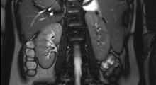
With the latest advances in Magnetic Resonance Imaging (MRI) technology, it is now possible to obtain high-quality MR images of the abdomen and pelvis that are equivalent to, and in many ways superior to, Computed Tomography (CT). MRI Enterography is an effective tool used specifically to image the small bowel to assess the presence and extent of Crohn’s disease and/or inflammatory bowel disease.
MR Enterography has been extensively used in patients with Crohn’s disease who require an accurate assessment of disease involvement and frequent follow-up studies. The main advantage of MR Enterography is the absence of ionizing radiation. In patients who have had numerous previous CT scans and in younger patients who are more radiosensitive, this study allows evaluation of the entire abdomen and pelvis without ionizing radiation exposure.
Preparation
No food or drink is to be taken for 8 hours before the scan so that your small bowel will be empty. An MRI Enterography requires that you drink approximately 48 ounces of a very dilute barium suspension over the course of 45 minutes before the actual scan at our Imaging Center. No radiation is involved with an MRI Enterography and the exam takes about 30-45 minutes to complete. An IV will be placed to give you medication and IV contrast during the exam.
During the Exam
Before the examination begins, you will be asked a series of questions about the presence of metal implants, such as artificial joints, or electronic devices, such as pacemakers because some of these may cause damage if they are put into the magnetic field of the MRI scanner. If so, please bring any documents with you to your appointment. If you have a heart pacemaker, please let CRL Imaging know at the time of scheduling your appointment. Unfortunately, you will not be able to have an MRI with a heart pacemaker and another test may be performed instead of an MRI.
The most important thing you can do to make the study successful is to keep as still as possible during the entire test, especially during the times you are asked to hold your breath. MRI may take several minutes, for the pictures to be taken. As with taking a picture of a moving object with an ordinary camera, blurry images result if you move during an MRI scan. You will have breaks during which you can breathe normally.
Follow-Up
After the procedure, you are able to return to a normal diet and activities immediately. As you may experience diarrhea, loose stools, and/or abdominal cramping, we encourage you to drink extra clear fluids for 24 hours after your procedure. This also helps remove the contrast from your system.
A board-certified Radiologist will interpret the scan and relate its information to the referring physician, who will in turn inform you of the results. All written reports will be available to the referring physician within 24 hours. Anytime immediate attention is needed, the referring physician will be contacted the day of the exam.