Virtual Colonoscopy
Seeing Inside
Colorectal cancer is the second leading cause of cancer deaths in the United States. Most colorectal cancers start as polyps – benign (non-cancerous) growths on the inner wall of the rectum and colon. Polyps develop slowly and can remain harmless for a long time before becoming aggressive cancer. Polyps, when detected, can be removed preventively.
Currently, doctors recommend that everyone be screened for colon cancer starting at age 50.
Virtual Colonoscopy (VC) allows for the evaluation of the entire surface of the colon for polyps and cancers.
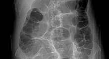
This technique in its entirety was first introduced in 1994 as an alternative to traditional colonoscopy. VC uses x rays and computers to produce two and three-dimensional images of the colon (large intestine) from the lowest part, the rectum, all the way to the lower end of the small intestine and display them on a screen. The procedure is used to diagnose colon and bowel disease, including polyps, diverticulosis, and cancer. VC can be performed with computed tomography (CT), previously called a CAT scan.
About the Procedure
VC is more comfortable than conventional colonoscopy for some people because it does not use a colonoscope. As a result, no sedation is needed, and you can return to your usual activities or go home after the procedure without the aid of another person. VC provides clearer, more detailed images than a conventional x ray using a barium enema, sometimes called a lower gastrointestinal (GI) series. It also takes less time than a conventional colonoscopy or a lower GI series.
Preparation
Because a clean and empty colon is necessary, you will be asked to prepare for the exam the day before it is scheduled.
The preparation consists of magnesium citrate and bisacodyl tablets the day before the exam and a bisacodyl suppository the morning of the exam.
Arrangements to receive the preparation will be made when the appointment is scheduled.
During the Procedure
A VC examination takes about 15-20 minutes and does not require sedation. During the procedure,
- The technologist will ask you to lie on your back on a table for the first portion of your exam.
- A thin tube will be inserted into your rectum, and air will be pumped through the tube to inflate the colon for better viewing.
- The table moves through the scanner to produce a series of two-dimensional cross-section images along the length of the colon. A computer assembles these images to create a three-dimensional picture that can be viewed on the computer.
- You will be asked to hold your breath during the scan to avoid motion artifact on the images.
- The scanning procedure is then repeated with you lying on your stomach.
After the Procedure
You may resume normal activities.
Follow-up
A board certified Radiologist from CRL Imaging will interpret the scan and provide a written report of the information to your referring physician within 24 hours.
Any finding needing immediate attention will be called to the referring physician the day of the exam.